|
EpiDerm-201TM
Under-developed Skin Model

주문은 최소 1개월 전에 하셔야 됩니다.
Features
- Partially Cornified Epidermal Structure
- Useful Tool to Study Modulators of Epidermal Differentiation
- 3 Days Younger than Standard EpiDerm (EPI-200)
- Differentiates into EPI-200
- Highly Reproducible
- Easily Handled Cell Culture Inserts
- NHEK-Based Skin Model
- Completely Serum-Free Media System
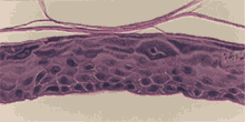
The EpiDerm-201 tissue model (EPI-201) is cultured in an identical manner to standard EpiDerm™ (EPI-200) except that the culture period is shortened by 3 days. The resulting EPI-201 tissue is thus less mature in terms of epidermal development. For example, histologically the spinous, granular, and stratum corneum layers are less developed than those in EPI-200 and the barrier function of EPI-201 is reduced. Because it is less developed, the EPI-201 tissue offers researchers the ability to investigate the effect of compounds designed to modulate earlier stages of epidermal differentiation.
Similar to EPI-200, the EPI-201 tissue consists of normal, human-derived epidermal keratinocytes (NHEK) which have been cultured to form a multilayered, metabolically and mitotically active, differentiating model of the human epidermis. Upon receipt, EPI-201 tissue can be returned to culture for an additional 3 days to produce a tissue which is nearly identical to standard EPI-200.
Various skin care and personal care companies are actively seeking alternatives to expensive, often highly-variable clinical testing. EPI-201 offers a cost-effective means to study modulators of epidermal differentiation or the effect of other active compounds on the skin. In addition, the EPI-201 tissue offers the advantage that specific epidermal phenomena (enzymatic activity, gene expression, cytokine production, etc) are more easily isolated and studied than in the clinical, more complex in vivo system.
Many of the standard protocols used for the EPI-200 system are easily adapted for use in the EPI-201 cultures. The use of cell culture inserts allows actives to be applied topically or into the medium (similar to sub-cutaneous administration). In addition, EpiDerm's rigid substrate make the tissue construct easy to handle and manipulate. Finally, EPI-201 (similar to EPI-200) is cultured in completely serum free medium, avoiding any undesirable interactions between test compounds and the numerous undefined components present in fetal bovine serum.
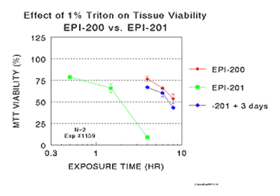
Histological cross-sections of EPI-201 tissues and their response to the positive control, 1% Triiton X-100, are shown in Figures 1-4. The effects of continuing EPI-201 tissues in various culture media are shown in Figure 5.
 |
|
Figure 1: Histological cross-section of EPI-201 tissue. Underdeveloped spinous, granular, and stratum corneum layers are apparent in comparison to those of normal human skin and the standard EPI-200 tissue shown in Figure 3. Final magnification = 440X.
|
 |
|
Figure 2: EPI-201 tissue, after storage for 24 hours at 4°C, was returned to culture for 3 days at 37°C and fed with 5.0 ml of EPI-201 differentiation medium (EPI-100-DM). Final magnification = 320X.
|
 |
|
Figure 3: Standard EPI-200 tissue grown in parallel with the EPI-201 tissue of Figure 2. Final magnification = 320X.
|
 |
|
Figure 4: Effect of Triton X-100 on the viability of tissue shown in Figures 1-3. 100 ml of 1% Triton X-100 were applied topically to the air-exposed surface of the tissues for specified time periods and MTT cell viability assays were run. The viability of the EPI-201 tissue was decreased to 50% of controls in 2 hrs while EPI-200 and re-cultured EPI-201 tissues still had 50 % viability after 7-9 hrs. These results imply that the barrier function of the EPI-201 is significantly decreased in comparison to that of EPI-200.
|
 |
|
Figure 5A. |
 |
|
Figure 5B. |
 |
|
Figure 5C. |
 |
|
Figure 5D.
Figure 5. Effect of culturing EPI-201 for 3 days with the following media:
A) Standard EPI-200 medium (used to produce EPI-200),
B) EPI-200 medium plus 5 x 10⁻⁹ M retinoic acid,
C) EPI-200 medium without growth factors and hormones, or
D) DMEM alone.
EPI-201 cultures were fed 5.0 ml of medium on Days 1 and 2 and fixed on Day 3. |
(See also EpiDerm Skin Model)
Technical Specifications
I. Ordering
- New Orders: MatTek Scientists will consult on your application at no additional charge. It is STRONGLY RECOMMENDED that you have this consultation BEFORE placing your first order to help ensure that all media and accessories needed to perform your application are included in the initial order.
- ALL Tissue Orders: Please allow 3-4 weeks from order date to delivery of tissues. Please contact MatTek Customer Service for additional details.
II. Cells
- Type: Normal human epidermal keratinocytes (NHEK).
- Genetic make-up: Single donor.
- Derived from: Neonatal-foreskin tissue.
- Alternatives: NHEK from adult breast tissue.
- Screened for: HIV, Hepatitis-B, Hepatitis-C, mycoplasma.
III. Medium
- Base medium: Dulbecco's Modified Eagle's Medium (DMEM).
- Growth factors/hormones: Epidermal growth factor, insulin, hydrocortisone and other proprietary stimulators of epidermal differentiation.
- Serum: None.
- Antibiotics: Gentamicin 5 µg/ml (10% of normal gentamicin level).
- Anti-fungal agent: Amphotericin B 0.25 µg/ml.
- pH Indicator: Phenol red.
- Other additives: Lipid precursors used to enhance epidermal barrier formation (proprietary).
- Alternatives: Phenol red-free, antibiotic-free, anti-fungal-free, or hydrocortisone-free medium and tissue are available. Agents are removed at least 3 days prior to shipment.
- Maintenance medium: EPI-201 kits are provided with differentiation medium (EPI-100-DM). After 3 days of culture using EPI-100-DM (5.0 ml changed every other day), EPI-200 tissues are produced from the EPI-201 tissues. Beyond that point, maintenance media usable for EPI-100 can be used (EPI-100 is a previous version of EPI-200). See EPI-200 technical specifications for more details.
IV. Tissue
- Kit: Under-developed EpiDermTM Skin Model (EPI-201) consists of 24 tissues which have been cultured for 3 days less than EPI-200.
- Substrate: Chemically modified, collagen-coated, 9 mm ID single well tissue culture plate inserts are used (e.g. Millicell CM, Nunc polycarbonate single well tissue culture plate inserts).
- Culture: At air liquid interface.
- Histology: 4-6 cell layers (basal, spinous, and granular layers).
- Stratum Corneum: <10 layers (based on TEM).
- Lot #'s: Tissue lots produced by each technician for each week are assigned a specific lot #. Typically, there are multiple lot #'s for any given week's tissue production. A letter of the alphabet is appended to the end of the lot # to differentiate between individual kits within a given lot of tissues. All tissue kits within a lot are identical in regards to cells, medium, handling, culture conditions, etc.
- Shipment: At 4°C on medium-supplemented, agarose gels in 24-well plate.
- Shipment day: Monday or Thursday.
- Lead Time: 3 weeks (for Monday shipment) maximum. Call Customer Service to see if shorter lead time is possible.
- Delivery: Tuesday morning via FedEx priority service (US). Outside US: Tuesday-Thursday depending on location.
- Shelf life: Including time in transit, tissues may be stored at 4°C for up to 6 days prior to use. However, extended storage periods are not recommended unless absolutely necessary. In addition, the best reproducibility will be obtained if tissues are used consistently on the same day, e.g. Tuesday afternoon or following overnight storage at 4°C (Wednesday morning).
- Length of experiments: EPI-201 cultures can be continued 3 days to produce EPI-200 tissues. After that, cultures can be continued for up to 3 weeks with good retention of normal epidermal morphology. Initially, EPI-201 cultures must be fed every other day with 5.0 ml of EPI-100-DM to produce EPI-200 tissue. After that point, the tissues are maintained in either long life maintenance medium (EPI-100-LLMM) or standard maintenance medium (EPI-100-MM). Cell culture inserts are placed atop washers (EPI-WSHR) or culture stands (MEL-STND) in 6-well plates to allow us of 5.0 ml.
- Alternative tissues:
EPI-201-PRF: Cultured using phenol red-free medium. All EPI-201 tissues are cultured for 3 days less than standard EPI-200 tissue.
EPI-201-AFAB: Cultured in antibiotic (gertamicin)-free and anti-fungal agent (Amphotericin B)-free medium. All EPI-201 tissues are cultured for 3 days less than standard EPI-200 tissue.
EPI-201-HCF: Cultured in hydrocortisone-free medium. All EPI-201 tissues are cultured for 3 days less than standard EPI-200 tissue.
EPI-200-1S: These undifferentiated tissues are grown for 1 day in submerged culture similar to the initial stages of standard EPI-200 production. These cultures are not exposed to the air-liquid inferface.
EPI-200-3S: These undifferentiated tissues are grown for 3 days in submerged culture similar to the initial stages of standard EPI-200 production. These cultures are not exposed to the air-liquid inferface.
EPI-200-5S: These undifferentiated tissues are grown for 5 days in submerged culture similar to the initial stages of standard EPI-200 production. These cultures are not exposed to the air-liquid inferface.
V. Quality Control and Sterility
- Visual inspection: All tissues are visually inspected and if physical imperfections are noted, tissues are rejected for shipment.
- End-use testing: EPI-201 tissues are continued in culture for 3 additional days and tested in an identical manner to EPI-200 tissue lots. The EPI-200 tissue is exposed to 1% Triton X-100 for 4, 6, 8, and 12.5 hours. The time of exposure required to reduce the tissue viability (ET-50) using the MTT assay is determined (See MatTek EpiDerm ET-50 protocol) for each lot of tissue. ET-50's must fall within the range of the 1996 EpiDerm database: 4.77-8.72 hours. ET-50's in customers' lab may differ slightly from the MatTek results.
- Sterility: All media used throughout the production process is checked for sterility. Maintenance medium is incubated with and without antibiotics for 1 week and checked for sterility. The agarose gel from the 24-well plate used for shipping is also incubated for 1 week and checked for any sign of contamination.
- Notification of lot failure: If a tissue lot fails our QC or sterility testing, the customer will be notified and the tissues will be replaced without charge. Because our QC and sterility testing is done post-shipment, notification will be made as soon as possible.
|