The EpiVaginal ™ Tissue Model
주문은 최소 1개월 전에 하셔야합니다.

Features
EpiVaginal Tissue Model
- Human 3-D Vaginal-Ectocervical Tissues
- Produced from Normal (Non-Transformed), Human-Derived Vaginal-Ectocervical Epithelial Cells
- Multi-Layered Tissues - Basal Layer and Multiple Non-Cornified Layers
- Two Tissue Series - VEC and VLC
- VLC Tissues - Contain Dendritic Cells, Infectable with HIV-1
- Ideal for toxicity studies of feminine hygiene, vaginal care, and microbicide products
- Useful for the Study of HIV-1, Other Sexually Transmitted Diseases (STDs)
- Serum-Free Medium Available
General MatTek Tissue Features
- Unsurpassed Long-Term Tissue Reproducibility
- 3-Dimensional, Highly Differentiated Tissues
- Metabolically, Mitotically Active Tissues
- Produced from Normal (Non-Transformed) Human Cells - Ideal for Genomics Studies
- Produced in Easily Handled Cell Culture Inserts
- Quantifiable, Objective Test Endpoints
- Cost Effective Alternative to Animal and Human Clinical Testing
- List of Contract Testing Labs Qualified to Run MatTek Tissue-Based Tests Available
Applications
- Microbicide Testing
- STD Infection
- Feminine Hygiene
- Inflammation
Data Sheet
To facilitate the study of vaginal-ectocervical (VEC) toxicity, pathologies, and basic mucosal phenomena, MatTek has developed the EpiVaginal series of tissue models. EpiVaginal tissues are based on normal, human-derived VEC epithelial and dendritic cells (DC). Four types of EpiVaginal are offered:
1) VEC-100 (Figure 1): An epithelial tissue containing epithelial VEC cells,
2) VLC-100 (Figure 1): A patented epithelial tissue containing epithelial VEC and immuno-competent dendritic cells,
3) VEC-100-FT (Figure 2): A full thickness version of VEC-100 which includes VEC epithelial cells and a fibroblast-containing lamina propria, and
4) VLC-100-FT (Figure 2): A patented immuno-competent version of the VEC-100-FT which includes dendritic cells.
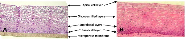
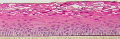
Figure 1: H&E stained histological (formalin fixed) cross-sections of A) VEC-100 in vitro reconstructed epithelial tissue models containing normal human VEC cells, and B) vaginal explant tissue. Both in vitro and in vivo tissues show nucleated basal and suprabasal cell layers followed by layers in which nuclei are lost and cells are filled with glycogen.
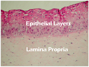
Figure 2: H&E stained histological (formalin fixed) cross sections of full-thickness EpiVaginal tissues. The VEC-100-FT tissues consist of VEC epithelial cells cultured atop a lamina propria (LP)-like collagen matrix that contains fibroblasts. The VLC-100-FT consists of VEC epithelial and dendritic cells in the epithelial layers and contains both fibroblasts and dendritic cells in the LP.
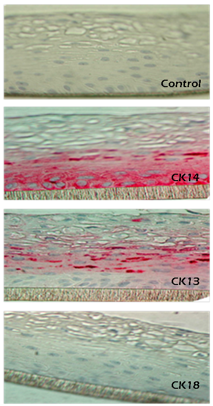
Figure 3: Formalin fixed cross-sections of the VEC-100 tissue immuno-stained for cytokeratins, CK13, CK14, & CK18. Basal cells are stained by CK14 and supra-basal cells by CK13; no cells are stained by CK18.
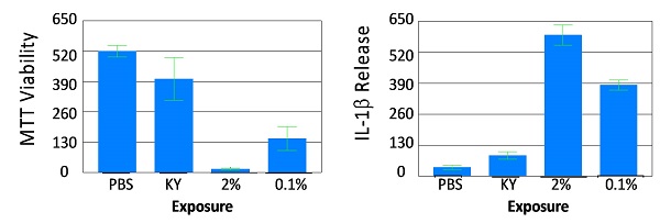
Figure 4: Viability (A) and Cytokine Release (B) of full-thickness vaginal ecto-cervical (VEC-100-FT) tissue model following exposure (18 hr) to formulations containing the common spermicide nonoxynol-9 (N9): a) PBS control and b) KY jelly (KY, 0% N9), c) KY N9 (2%), and d) N9 (0.1%). As viability of the tissue decreases, the IL-1β increases.
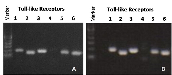
Figure 6: RT PCR showing Toll-like Receptor (TLR) expression for: A) organotypic vaginal-ectocervical (VEC) tissue model and B) Human cervical tissue explant.
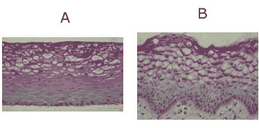
Figure 7: PAS Staining for Glycogen. Photo-micrographs of PAS reacted cross-sections of: A) reconstructed vaginal ectocervical tissue (VEC-100) and B) excised human vaginal tissue. Counter stain = hematoxylin. Glycogen shows up reddish-pink. In both tissues, the intensity of glycogen staining increases as the apical surface is approached. Note: In vivo, glycogen is released by the epithelial cells and fermented to lactic acid by lactobacilli (i.e. Doderlein's bacilli) that are part of the normal vaginal flora, accounting for the mildly acidic pH of vaginal fluid. Click on photo to see larger image.
The EpiVaginal tissue models exhibit in vivo-like morphological and growth characteristics which are uniform and highly reproducible. EpiVaginal is a multilayered tissue consisting of an organized basal layer and multiple non-cornified layers analogous to native human vaginal-ectocervical tissue . The tissue expresses cytokeratin K14 in the basal and supra basal layers and cytokeratin K13 in the suprabasal tissue layers.
Various industrial and toxicology laboratories are actively seeking alternatives to expensive clinical or whole animal testing. The protocols for using EpiVaginal are clear and straightforward. Feminine hygiene, personal care, and pharmaceutical companies have initiated in vitro toxicology testing to evaluate their raw materials and final product formulations.
Table 1: EpiVaginal ET-50 ranges for various feminine hygiene products
|
Material Type |
ET-50 Range |
|
Triton X-100 (1%)
|
0.75-1.75 hr
|
|
Feminine washes (3 tested)
|
1-3 hrs
|
|
Spermicides (4 tested)
|
3-7 hrs
|
|
Anti-itch creams (4 tested)
|
6-18 hrs
|
|
Lubricant (2 tested)
|
>24 hrs
|
|
Antifungal products (3 tested)
|
>24 hrs
|
|
Douche (2 tested)
|
>24 hrs
|
Method: Duplicate VEC-100 tissues were treated with 83 uL of various feminine hygiene products for a range of exposure times. Following exposure, the product was washed from the VEC-100 tissue and the viability of the VEC-100 tissue was determined using the MTT assay. The exposure time that reduced the tissue viability to 50% (ET-50) was determined by interpolating between exposure times which bracketed 50% viability. A lower ET-50 indicates a more aggressive, more irritating product.
Straight forward protocols are available for harvesting RNA to analyze gene expression or for measuring cytokines released into the culture medium (analyzed using ELISA assays). Companies and researchers utilize antibiotic/antifungal free EpiVaginal tissue (VEC-100-AFAB) to grow various opportunistic infections and pathogenic microbes in order to study their effects on the vaginal tissues. In addition, the VLC models are infectable with HIV-1 (Figure 6) and can be used to study HIV infection, transmission, and microbicides intended to prevent heterosexual passage of HIV. Finally, the tissue is amply suited to study a broad variety of pathogens that invade the vaginal-ectocervical environment along with prophylactic remedies there to.
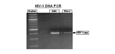
Figure 8: HIV-1 infection of VLC-100 tissue following topical exposure (24hr) to 60,000 CPM of HIV-1 ADA or NL4-3 virus. After exposure, tissues were washed 3X and then cultured for an additional 24 hr. Cellular DNA was then extracted and HIV-1 transcripts were detected using HIV-1 gag specific primer pairs. Click on photo to see larger image.
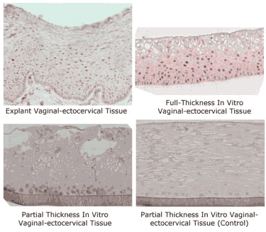
Figure 9: Micrographs showing nuclear staining of estradiol receptor (red) on: 1) Explant vaginal-ectocervical tissue (positive control), 2) Full-Thickness vaginal-ectocervical tissue model (VEC-FT), 3) partial-thickness vaginal-ectocervical (VEC) tissue, and 4) VEC tissue as negative control (without antibody). Formalin fixed tissues were labeled with rabbit anti-estrogen receptor (1:20 dilution) (Zymed Laboratories, Inc, South San Francisco, CA), incubated for 2 hr at room temperature, washed, and color was developed using alkaline phosphatase with Fast Red as substrate. Click on photo to see larger image.
Microbicides Testing - EpiVaginal™
Infection through the vaginal-ectocervical (VEC) tissue is believed to be the main route for the heterosexual transmission of the human immunodeficiency virus (HIV) in women.
Recently, a tissue culture-based model of the VEC (EpiVaginal) has been developed. Normal, human VEC epithelial and dendritic cell co-cultures were used to form a three-dimensional tissue using specially formulated medium. The in vitro engineered tissue reproduces many of the histological and ultrastructural features including basal, parabasal, glycogenated intermediate, and the superficial cell layers.
Preliminary experiments showed the use of this tissue model and the MTT tissue viability assay for predicting VEC irritation of microbicides*. Different concentrations of Nonoxynol-9 (N-9), carrageenin-I, and methyl cellulose were dosed topically and the viability of the VEC tissue was determine by MTT.
Following 24 hr exposure to N-9 (0.1%) tissue viability was reduced to 51%. In contrast, carrageenin-I (20%) reduces viability to 77% and no effect was observed for methyl cellulose (up to 20%). H & E staining showed irritation of epithelial lining following N-9 treatment greater than or equal to 0.1%. Experiments also showed that HIV virions do not pass freely through the tissue but they infect cells in the reconstructed VEC tissue model containing dendritic cells.
In conclusion, the tissue model is likely to serve as a useful tool to screen new or existing microbicide formulations for vaginal toxicity and microbicidal efficacy.
====================
Excerpted from, "HUMAN VAGINAL-ECTOCERVICAL TISSUE MODEL FOR MICROBICIDE IRRITATION STUDIES" (MatTek Corp. Technical Reference TR-320) - Presented at "Microbicides 2004", London, England, March 28-31, (2004).


